Research Digest
|
Therapy and Rehabilitation
Unveiling the Expert Eye: How Eye Tracking is Revolutionizing Ultrasound-Guided Embryo Transfer
Medical & Healthcare
Expertise & Performance

Video: Recording made with Neon during a simulated ultrasound-guided embryo transfer procedure. The surgeon’s gaze follows the tip of the catheter (visible as the white blob on the ultrasound image) as it advances into the cervical canal. Video courtesy of Dr. Josselin Gautier.
Infertility affects an estimated 1 in 6 people globally, and In Vitro Fertilization (IVF) remains one of the most effective assisted reproductive technologies, but outcomes still vary significantly. A key step in IVF is Embryo Transfer (ET), a delicate procedure in which an embryo is placed into the uterus. Despite its critical importance, success rates depend heavily on the skill of the practitioner.
Until now, assessing the visual strategies behind this skill-intensive procedure has been a challenge. In the first study of its kind, Josselin Gautier and colleagues from the Laboratoire Traitement du Signal et de l'Image (LTSI) of the University of Rennes (France) captured eye movement data during ultrasound-guided embryo transfer (UGET) to better understand what sets expert practitioners apart.
A New Approach: Eye Tracking for Precision Medicine
Using Pupil Labs Neon eye tracking system, researchers recorded the gaze behavior of Assisted Reproductive Technologies (ART) specialists as they performed UGET on a Gynos Virtamed simulator. The simulation allowed for realistic head-unrestrained movement in dim-light conditions, mimicking actual clinical environments.
Eye movements were recorded across all three stages of the procedure:
Cervical canal navigation
Uterine navigation and embryo deposition
Catheter removal
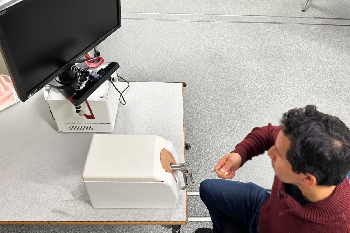
Figure 1: Equipment used in the study, including the Guardia catheter system, one of the uterus models, and the Gynos simulator setup with screen and controls. Image courtesy of Dr. Josselin Gautier.
Key Findings and Insights
This innovative approach revealed distinct visual strategies and expertise-linked patterns that could help shape the future of medical training.
Eye tracking in complex medical settings is feasible. The study successfully demonstrated that portable eye trackers like Neon can reliably record eye positions over multiple objects in a complex medical scene and within small areas of interest on an ultrasound screen. Practitioners reported no discomfort wearing the headset during the procedure.
Experts and novices show different gaze strategies. Some practitioners adopted a target-based strategy, focusing primarily on the ultrasound image, a pattern typical of experts in image-guided procedures. Others showed a switching/tool-following approach, alternating more between screen, tools, and model, a behavior often seen in less experienced users.
More experience, fewer saccades ART specialists with higher expertise exhibited longer fixation durations and lower saccade rates, indicating a more deliberate and efficient visual approach.
Surprising pupil dilation in experts Contrary to expectations, more experienced practitioners showed greater pupil dilation during the task. While pupil dilation typically correlates with cognitive load, this may instead reflect high motor precision demands, rather than stress or inexperience.
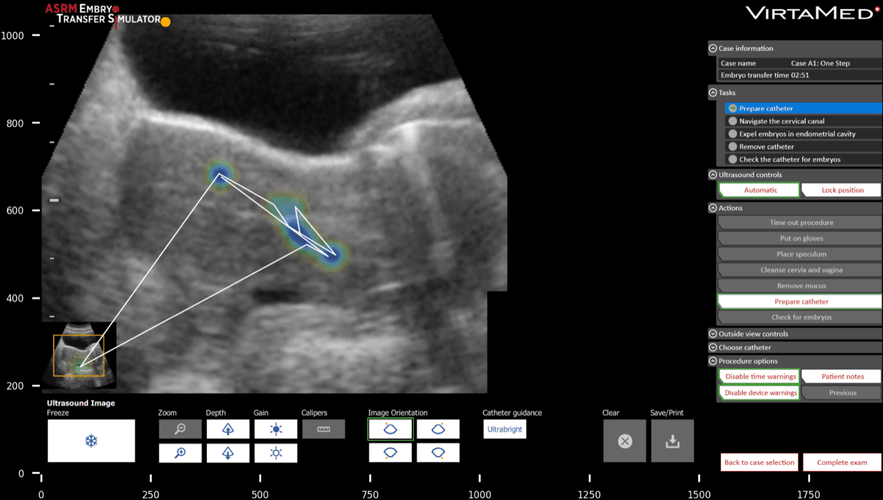
Figure 2: Ultrasound image of the Gynos simulator with an overlaid heatmap and scanpath, showing where the surgeon focused their gaze throughout the procedure. Image courtesy of Dr. Josselin Gautier
Implications for Medical Training
This study represents a significant step forward in medical skill assessment and training for ultrasound-guided procedures. By combining simulation-based training with eye tracking, it becomes possible to:
Identify objective markers of expertise
Distinguish between novice and expert visual strategies
Develop tailored training programs that foster expert-like gaze behavior
As IVF procedures continue to evolve, integrating tools like Neon into simulation-based training could help clinicians refine their skills, reduce variability in outcomes, and ultimately improve patient care.